
Short answer
On the post-insertion chest X-ray (CXR), trace the catheter’s course, check its tip position and alignment, and look for pneumothorax or haemothorax. For adult upper-body central venous catheters (CVCs), suitable tip regions include the lower superior vena cava, cavo-atrial junction or high right atrium, depending on the device and purpose. A satisfactory-looking CXR does not, by itself, prove that a catheter is venous.[1][2]
What does the CXR add?
Ultrasound, along with pressure transduction and blood-gas analysis during insertion confirms needle and guidewire placement in the accessible vein. It does not necessarily show where the catheter finally travels or where its tip ends. The CXR adds a view of the intrathoracic course and tip, and can reveal pleural complications.[1]
Keep three questions separate: is the catheter venous, is its tip appropriately positioned, and has insertion caused a complication? Transduce the CVC and confirm an appropriate venous waveform and pressure before use, as described in the internal jugular CVC insertion guide. CXR review complements that check.[3]
Where should the tip lie?
The superior vena cava (SVC) descends on the right of the mediastinum and joins the right atrium at the cavo-atrial junction. The catheter should lie along the vessel, with its tip parallel to the vessel wall. A tip that points sharply into the wall is concerning even if its apparent depth seems reasonable.[1]
For upper-body catheters, the preferred tip regions are the lower third of the SVC, the cavo-atrial junction and the high right atrium. This does not make every right atrial position acceptable: a deeply intracardiac catheter is different from a tip just beyond the junction. Apply the catheter’s instructions and the intended treatment when deciding whether its position is suitable.[1]
Using the carina as a landmark
Identify the carina, where the trachea divides into the main bronchi. The cavo-atrial junction is not directly visible on a plain CXR, but its level can be estimated at approximately two vertebral body units, or about 4 cm, below the carina. Treat this as an anatomical guide rather than an exact measurement for every patient.[1]
The carina itself is not the intended tip target. Assess depth together with course and alignment, particularly for a left-sided line that must turn from the left brachiocephalic vein into the SVC. Patient position and respiration can also change tip position.[1]
Read the CXR systematically
A right internal jugular line usually descends towards the right-sided SVC. A left internal jugular or left subclavian line normally crosses the upper mediastinum through the left brachiocephalic vein before turning down into the SVC. Trace the whole visible course rather than judging the tip in isolation.[4]
Which appearances need further assessment?
| Finding | Concern |
|---|---|
| Tip turns up into the neck or travels laterally away from the SVC | Entry into an unintended vein |
| Catheter loops or doubles back | Malposition or kinking |
| Tip hooks near the right tracheobronchial angle | Possible azygos placement; a frontal CXR may not settle this |
| Catheter descends on the left of the mediastinum | An abnormal route, including arterial placement or a persistent left SVC; the image alone cannot decide which |
| Tip points sharply into the vessel wall, or extends deeply into the heart | Risk of vessel or cardiac injury; reassess suitability before use |
These appearances are reasons to investigate, rather than definitive diagnoses from a single projection.[4]
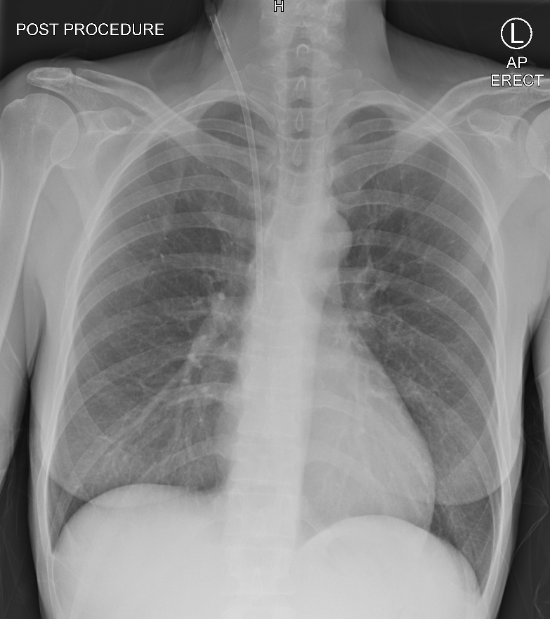
In this example, a right internal jugular CVC turns laterally into the right axillary vein rather than descending into the SVC. This illustrates why the whole catheter course matters.[5]
What can a reassuring CXR miss?
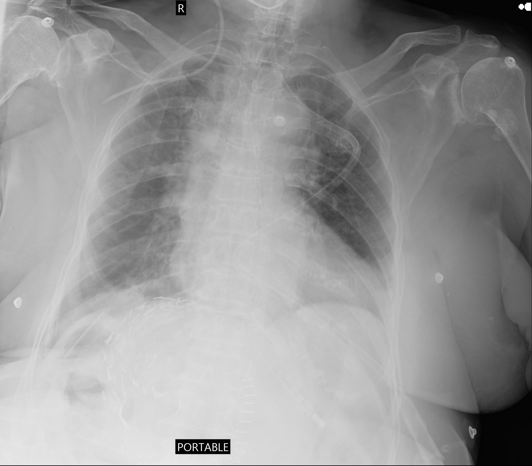
A frontal CXR is a two-dimensional projection. Vessels and other structures overlap, so a catheter projected over the SVC may still be arterial or outside the vein. Resolve discrepancies between the image, waveform and clinical findings before use.[2]
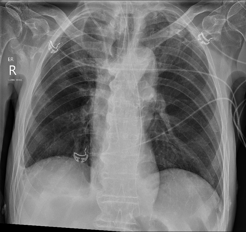
A small or developing pneumothorax may be absent from the initial image. New breathlessness, chest pain, hypoxaemia or haemodynamic deterioration still needs urgent assessment after an apparently satisfactory CXR. Use further bedside assessment and imaging as clinically indicated; do not wait for a routine report in a deteriorating patient.[2]
When can the line be used?
For the internal jugular insertion pathway in this handbook, obtain and review a CXR before routine use, alongside confirmation of venous placement and lumen function. Document the catheter course, tip position, any complication, and the decision about use. If adjustment or replacement is required, arrange it with an experienced clinician and reconfirm placement before routine use.
If treatment cannot safely wait for the CXR, follow the urgent-use exception in the insertion guide: confirm venous placement, assess for immediate complications, and document the responsible clinician’s decision and rationale. Arrange prompt CXR and review. Urgency does not make uncertain venous placement acceptable.[1][3]
References
-
Johnston AJ, et al. Association of Anaesthetists guidelines: safe vascular access 2025. Anaesthesia. 2025;80:1381–1396. doi:10.1111/anae.16727.
-
Bodenham A, et al. Association of Anaesthetists of Great Britain and Ireland: safe vascular access 2016. Anaesthesia. 2016;71:573–585. Referenced for imaging limitations and recognition of insertion complications.
-
Faculty of Intensive Care Medicine and Intensive Care Society. Invasive procedure safety checklist: CVC insertion. 2023.
-
Gibson F, Bodenham A. Misplaced central venous catheters: applied anatomy and practical management. Br J Anaesth. 2013;110:333–346.
-
Singh R. Misplaced central venous line. Case study, Radiopaedia.org. rID: 212475. Published 31 May 2025.
Cited at: 1
-
Ängeby E, et al. Central venous catheter tip misplacement: a multicentre cohort study of 8556 thoracocervical central venous catheterisations. Acta Anaesthesiol Scand. 2024;68:520–529.
Cited at: 1